Facility Overview
The Imaging Core suite in OMRF’s research tower houses sensitive instruments in dedicated rooms built over a vibration-damping sub-floor (designed by Chad Himmel, P.E., of JEAcoustics). There is also optimized space for sample preparation, tissue sectioning and off-line computing. Sample preparation equipment includes two automated tissue processors, a paraffin embedding station, four microtomes, two cryostats, and an automatic coverslipper. Imaging Core instruments include a cryo-ultramicrotome, a high-pressure freezer, slam freezers, freeze fracture equipment, a freeze substitution system and a sputter coater, as well as
- Standard upright and inverted microscopes
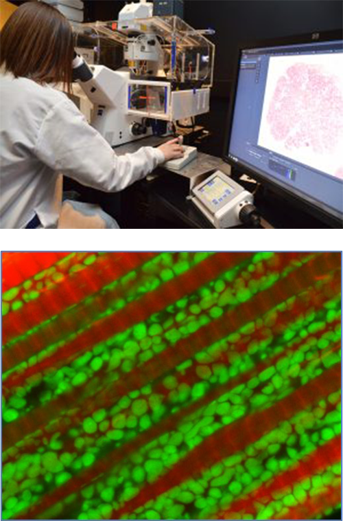
- Three Zeiss LSM-based laser scanning confocal microscopes (a LSM510, LSM 710 and a LSM 880 (all with spectral unmixing capabilities and environmental chambers for live cell work). The 880 is equipped with an Airyscan detector capable of live-cell super-resolution imaging
- An Olympus FV 1000 confocal microscope with incubation for live-cell imaging
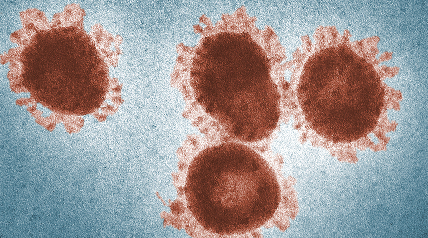
- Hitachi H-7600 transmission electron microscope fitted with a Kodak 4Kx4K 4.0 camera
- Structured illumination microscope with Colibri LED illumination
- A Zeiss Axiosscan 7 whole slide scanner. This system is capable of storing 100 slides at a time and image using brightfield, fluorescent, and polarized modalities
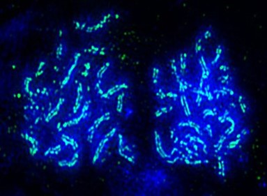
The core also houses a super-resolution DeltaVision OMX-SR microscope. As the only super-resolution microscope in Oklahoma, this instrument provides fluorescent microscopy images with much higher resolution than is possible in conventional fluorescence microscopy through two methods: structured illumination microscopy or SIM, and single molecule localization microscopy or localization microscopy. These methods effectively double or quadruple the resolution of conventional microscopy and are applicable to a wide range of research approaches. Computer support includes a 3D deconvolution 1.6TB data server and a dual zeon 500 GB data server. Also available are 3D deconvolution and data analysis workstations and extremely high-resolution printers.
The core has recently begun offering Spatial Transcriptomics services using both the Nanostring (GeoMx DSP) and 10X Genomics (Visium) platforms. These platforms allow investigators to combine histological or immunohistological information with sensitive high-plex profiling of protein or RNA transcripts at the cellular level, allowing for spatial resolution of functionally distinct cell populations and structures within the morphological context of the tissue. The systems use paraffin embedded or frozen tissue sections and fluorescent or chromogenic marker overlays to define the structural morphology of the tissue and help identify cell types or regions of interest for deeper examination. The imaging core is capable of processing the samples from tissue collection, through staining and library preparation, to sequencing.
Imaging Core Leadership
Gary Gorbsky PhD serves as the Director of the Imaging Core. Dr. Gorbsky is the Chair of the Cell Cycle and Cancer Biology Program at OMRF, has been an acting officer of the American Society of Cell Biology, and is an expert microscopist. He will have primary administrative and scientific oversight of the Imaging Core. Dr. Gorbsky is available to COBRE Investigators to provide advice on the proper design, implementation, and interpretation of imaging experiments.
Ben Fowler is the Imaging Core Manager and oversees all aspects to the daily operations of the Imaging Core. Mr. Fowler holds dual Master of Science degrees in Microbiology and Cell Biology. He has been managing the Imaging Core since its inception in 1999. Mr. Fowler has extensive experience in developing experimental protocols for fluorescence and electron microscopy, maintaining the full range of equipment within the Imaging Core, and in managing the personnel and business aspects of the facility.
Training
Mr. Fowler and two of the other full-time technical staff in the facility have extensive experience in training new users to utilize the full suite of light and electron microscopy equipment, following teaching protocols that have been developed over the past ten years. Access to each instrument is controlled by key-card activated doors and log-in requirements for the operating software.
Maintenance
The microscopes are covered by service contracts and Mr. Fowler performs preventative maintenance and quality control procedures as needed. Mr. Flower and his staff are able to perform common minor repairs so that down time is minimized.
Organization
Mr Fowler provides daily management of the Imaging Core while Dr. Gorbsky provides administrative oversight. Prospective users are trained in the safe and effective use of individual microscopes by Core technicians. Use of the instruments is monitored by the appropriate Core personnel until the user is approved to operate the instrument independently, including after hours.
Time allocation
User reservations are regulated by an automated, web-based calendar system which is monitored throughout normal working hours by Core personnel.
